Melody Alsaker's Research to Revolutionize Medical Imaging & Save Lives
This year, Melody Alsaker, Alphonse A. & Geraldine F. Arnold Distinguished Professor and associate professor of mathematics at Gonzaga, has been places that would stir awe in her younger self. From organizing a conference session on her research specialty in Tokyo to deeper collaborations between colleagues in Helsinki, Alsaker has set the standard for global scholarship in her field.
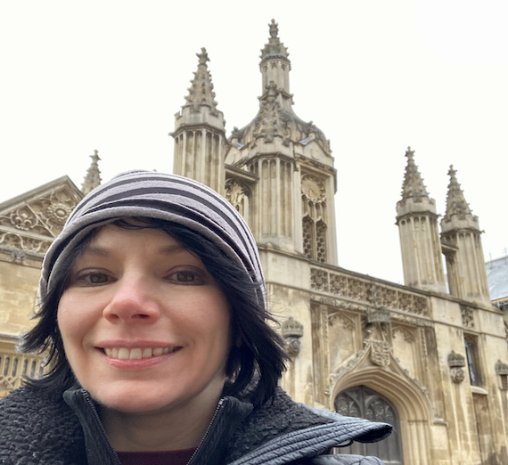
Just this spring, the researcher was invited to speak at a workshop at Cambridge University. Her session with peers and experts in her field focused on the mathematics of medical imaging and electrical impedance tomography (EIT), her area of research. EIT is a relatively new technology that uses electrical signals to create human body images. Unlike other types of medical imaging, such as X-rays or CT scans, EIT doesn't expose patients to harmful radiation. It is a much more portable and cost-effective technology that can be taken in an ambulance, helicopter, or battlefield.

There was just this sense of being a part of this rich tradition and all this history, and I'm just making my small contribution.
While at the Isaac Newton Institute, she marveled at the glass protection of a chalkboard containing the work of mathematician Andrew Wiles, who famously proved a long-unsolved theorem. Many other spaces were off-limits, but being with her peers, delivering a report of her research, and absorbing the rich history of her discipline made it worth it.
For Alsaker, her time at Cambridge was hard-earned and well-deserved.
Alsaker's success is grounded in a commitment to improving EIT technology, growth, and dedication. Her diligence and belief propelled her to college as a non-traditional student at age 23 with a GED, then to her master's, and finally, her Ph.D. All this work was undertaken after growing up in a household with shifting priorities – survival was the main objective. Even with a childhood home that couldn't emphasize education, Alsaker was able to tie together a robust academic experience that serves as a model for students and faculty from non-traditional backgrounds. With her latest lecture at Cambridge, she becomes part of a rich educational tradition, which improves life-saving technology, and shows the way for aspiring mathematicians.
How Alsaker's research improves machine vision technology
EIT aims to produce images that are good enough to be medically valuable and save resources as a low-cost option. An EIT machine is a small, less-expensive device, costing tens of thousands of dollars versus a million-plus for MRI/CT scanners. The challenge is that the images are still low-resolution, and this barrier is tangled in the hard math behind the imaging. Converting the electrical measurements into a viable image requires refining a complicated math problem. .
While fine-tuning of imaging is still in the works, the COVID-19 pandemic necessitated more reliance on EIT imaging, particularly ventilation monitoring. In this case, EIT primarily visualizes the air moving in and out of the lungs. Watch this fascinating process in the video below.
In an unfortunate twist of fate, Alsaker witnessed the technology's intersection with a real-world patient's fate – her father. When her father was put on ventilation, it was too late, and he passed away later that day. His passing and the complications around mortality, COVID, and lung health aroused complex thoughts in Alsaker but also ushered in a renewed commitment to her research.
"When it happened, I was feeling like, this is why I keep working on this,” she said, “because I think that what I do has the potential to impact people in the future. And I’m just making a small contribution to the overall body of research. But I do what I can.”
Bringing it back to Cambridge

At Cambridge, her presentation focused on artificial neural networks trained to understand the contours of human organs to generate missing parts and boundaries of imaging, which has not been done before. This network helps to shore up missing elements lost to the visual noise during the imagining process. But how does a neural network detect abnormalities? Alsaker says it goes back to training the network to detect specific abnormalities, with different neural networks detecting pathologies. Sometimes, irregularities are known more markedly, including cancerous tumors, which conduct more electricity than benign tumors.
Her work also has connections to "computer vision," or whether computers interpret images meaningfully. Can it give us information about the quality of an image? A "phantom" or user-generated image for research is a created image that one uses for its numerical data for testing purposes – algorithms offer pixel-to-pixel comparisons. However sophisticated and rigorous these technologies are, they still cannot beat the human eye. What Alsaker describes and the technology she is developing in partnerships with others is set on the bleeding edge where significant changes regarding machine vision and its abilities may have the chance to catch up to the human eye.
As an associate professor of mathematics at Gonzaga who is welcomed as an expert at higher education institutions like Cambridge University, Alsaker offers students and the world a chance to "see" what humanity can accomplish through her and others' aptitude and innovations.